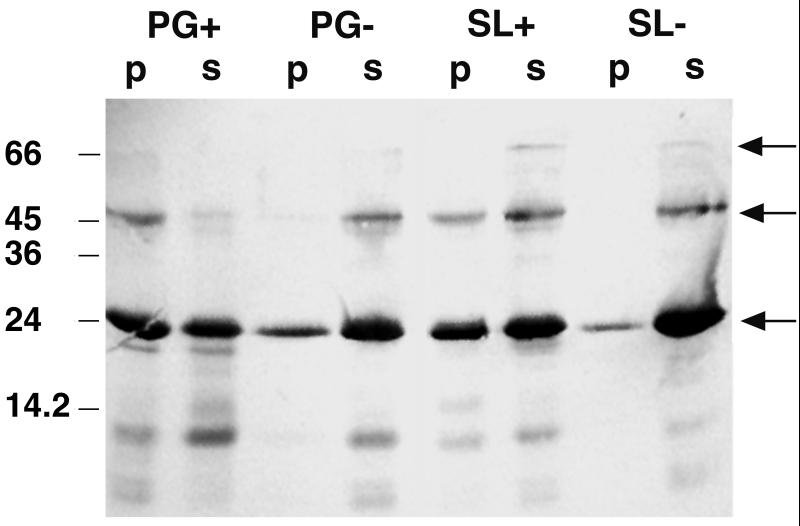
FIG. 3.
Western blot showing the affinity of the SLH polypeptide to itself and to isolated cell wall components. Ten micrograms of the SLH polypeptide and 0.5 mg of the cell wall component in a total volume of 100 μl were used in the interaction study. Twenty microliters of each fraction (p and s) was applied to an SDS gel. The blot was prepared with antibodies against the SLH polypeptide. Fractions p and s represent bound and unbound proteins, respectively. Arrows indicate bands representing the SLH polypeptide monomer (23 kDa), dimer, and trimer.

