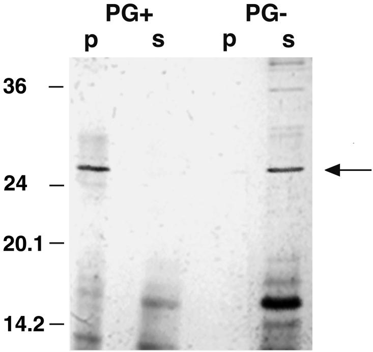
FIG. 6.
Interaction of an N-terminal cleavage fragment of the S-layer protein with native peptidoglycan-containing cell wall sacculi (PG+). A silver-stained SDS gel is shown. Fractions containing 20 μg of the cleavage fragments and 0.5 mg of the cell wall components were used (total volume, 100 μl), and 20 μl of each sample was applied to the gel. Bands representing the N-terminal cleavage fragment of the S-layer protein (27 kDa) are indicated by an arrow. p, fraction containing protein bound to the corresponding cell wall component; s, unbound protein. The migrations of marker proteins (in kilodaltons) are indicated on the left.

