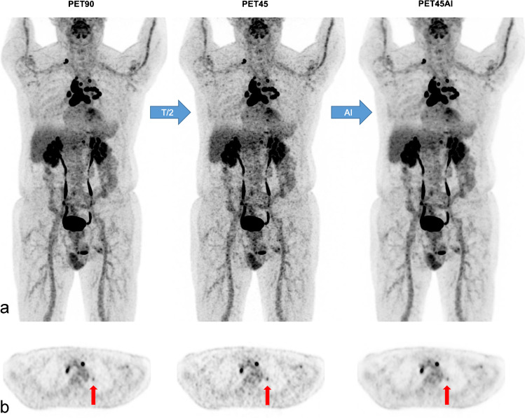
Fig. 3.
Concordant lesions A 77-year-old man (78 kg; BMI 24 kg/m2) with multifocal lymphadenopathy of unknown origin. MIP views (a) and axial PET slices (b) of [18F]FDG PET90, PET45, and PET45AI. Detection of small left suprahilar lymphadenopathy in all PET series (vertical arrows in b) with respective standard SULmax of 1.8 (PET90), 2.3 (PET45), and 1.7 g/ml (PET45AI). Nonetheless, PET45 images are noisier than PET90 or PET45AI images, particularly in the liver

