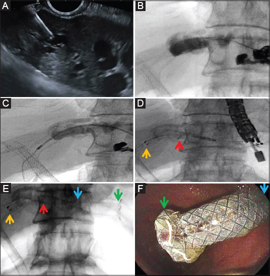
Figure 1.

Endoscopic ultrasound-guided hepatico-gastrostomy. (A) Transgastric, transhepatic puncture of a dilated left biliary duct with a 19-G fine-needle aspiration needle under endoscopic ultrasound guidance in a patient with Klatskin tumor and neoplastic ingrowth of previously placed percutaneous metal stents. (B) Contrast injection showing correct biliary access and intrastent neoplastic ingrowth, followed by guidewire cannulation of the biliary duct. (C) Tract creation through a 6-Fr cystotome and guidewire redirected inside the former metal stent. (D) Initial deployment of a partially covered metal stent with the uncovered portion inside the biliary tree and the previous stent, whilst the covered portion is deployed transhepatic and transgastric: yellow arrow, extremity of the stent (uncovered portion); red arrow, transition between uncovered and covered portion of the stent. (E) Final deployment of the stent: yellow arrow, intrabiliary extremity; red arrow, transition between uncovered (endobiliary) and covered (transhepatic) portion; blue arrow, transition between the transhepatic and transgastric portion; green arrow, intragastric extremity with antimigration flange. (F) Endoscopic view of the intragastric end of the hepatico-gastrostomy stent
