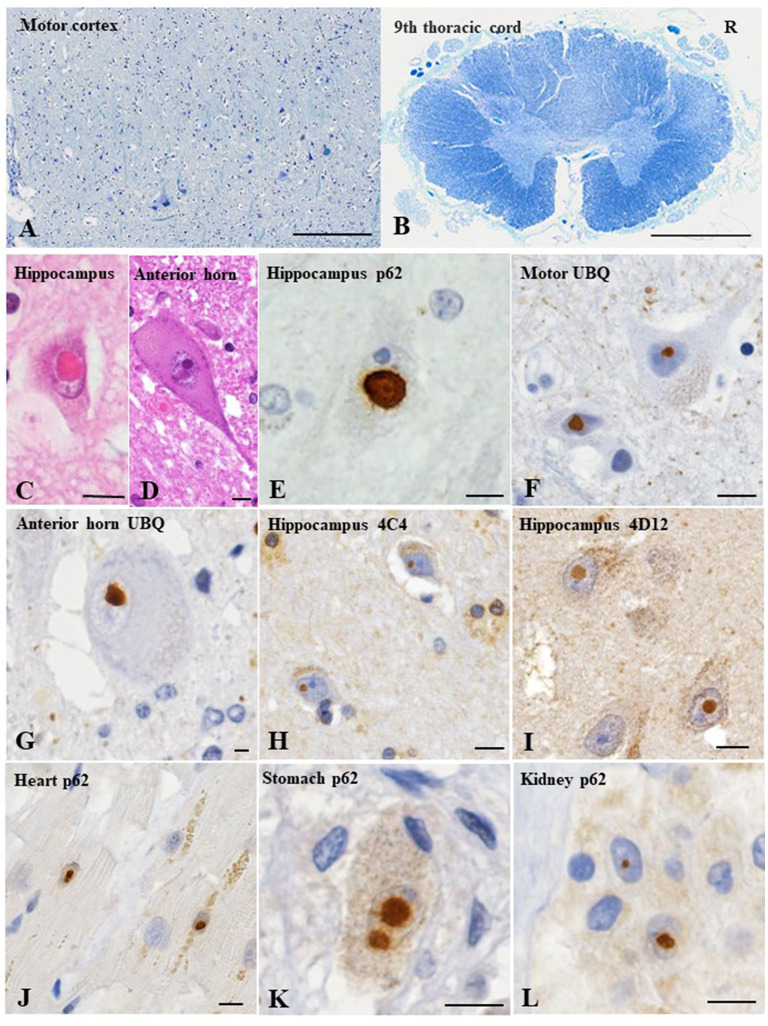
Figure 2.
Pathological findings of the present case. The motor cortex shows slight loss of neurons (A). Thoracic spinal cord shows myelin pallor of the lateral corticospinal tract bilaterally, more apparent on the right side (R), reflecting the left thalamic hemorrhage (B). Hematoxylin and eosin staining reveals an eosinophilic, round, neuronal intranuclear inclusion in the hippocampus (C) and anterior horn of the lumbar cord (D). Immunohistochemical evaluations reveal ubiquitin- and p62-immunopositive neuronal intranuclear inclusions in the hippocampus (E), motor cortex (F) and spinal anterior horn (G). In addition, uN2CpolyG immunoreactivity is confirmed by two specific antibodies (4C4 and 4D12) (H, I). p62-positive neuronal intranuclear inclusions are evident in the cardiac muscle cells (J), Auerbach's plexus of the stomach (K), and renal tubule epithelial cells (L). Klüver–Barrera staining (A, B); p62 (E, J–L); ubiquitin (F, G); uN2CpolyG (4C4) (H); uN2CpolyG (4D12) (I). Bars = 100 μm (A), 2 mm (B), and 10 μm (C–L).

