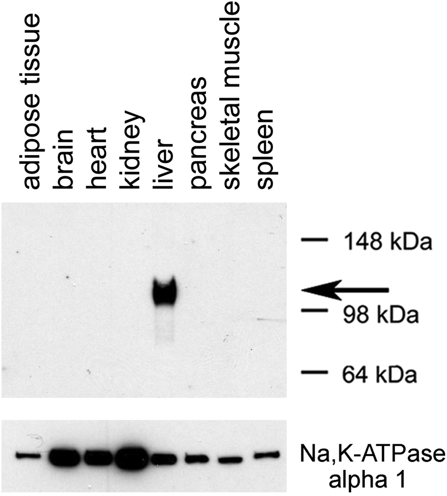
Fig. 6.

Selective expression of Dq in the liver of a mouse treated with the AAV-TBG-Dq virus. The AAV-TBG-Dq virus was injected into the tail vein of a C57BL/6 mouse. Three weeks later, membranes were prepared from the indicated tissues. Subsequently, membrane extracts were subjected to SDS-PAGE under reducing conditions (in the presence of 10% β-mercaptoethanol). Dq expression was detected via Western blotting using a rabbit monoclonal anti-HA tag antibody (note that Dq contains an N-terminal HA tag; see Fig. 5). The arrow indicates the position of the designer receptor (Dq). For control purposes, the blot was also probed with a mouse anti-α1 sodium potassium ATPase monoclonal antibody (predicted molecular mass: 112 kDa).
