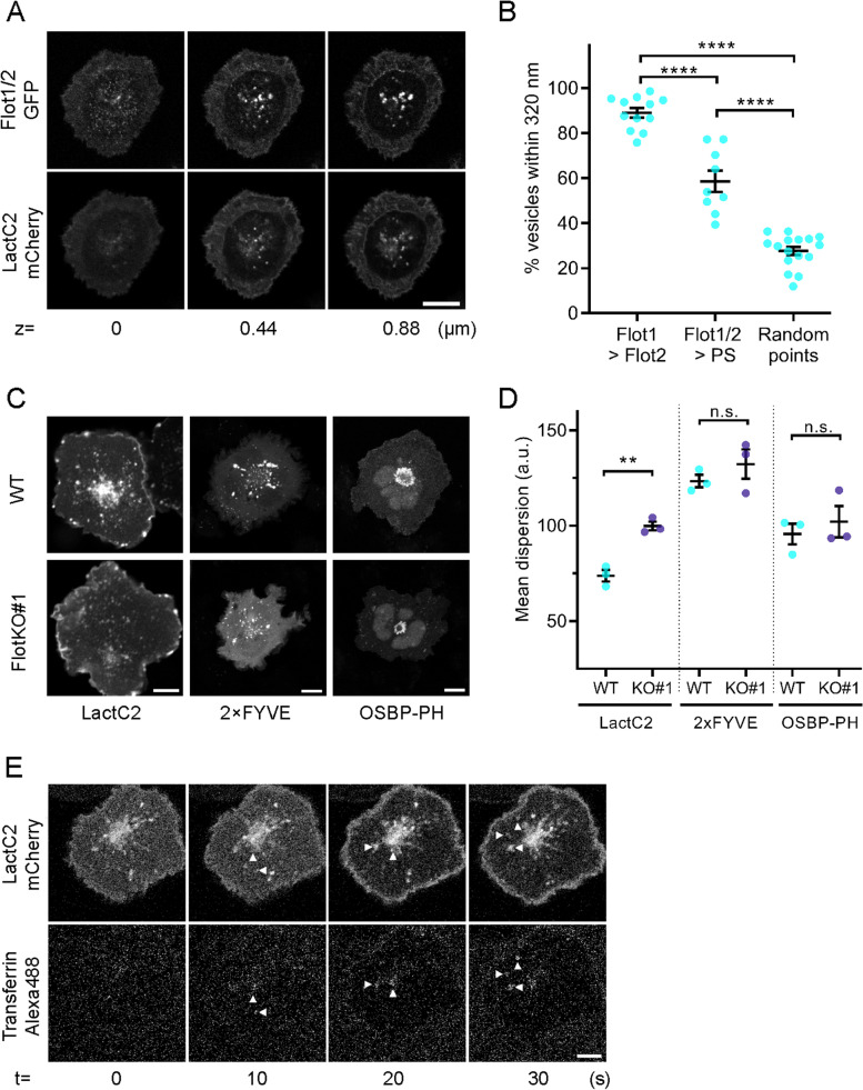
Fig. 4.
Fast transferrin-TfR recycling axis is demarked by phosphatidylserine. A Representative confocal images at 3 different z-levels at (0 μm) and above (0.44 μm and 0.88 μm) the immunological synapse of activated Jurkat T cells expressing Flot1/2-GFP and the PS-sensor LactC2-mCherry. B Nearest neighbour analysis between green and red channel identifying % of vesicles positive for both colours (≤ 320 nm apart; % Flot1 vesicles also containing Flot2, % Flot1/2 vesicles positive for LactC2 (PS) and random points to each other) in GFP and mCherry as described in [15]. C Representative confocal images of fixed, 20-min activated WT (upper) or FlotKO (lower) Jurkat T cells expressing PS sensor LactC2-GFP, PI(3)P-sensor 2 × FYVE-GFP or PI(4)P-sensor OSBP-PH-GFP. Scale bar = 5 μm. D Mean fluorescence dispersion of GFP channel in WT and FlotKO Jurkat T cells. E Representative time series of confocal imaging of an activated Jurkat T cell expressing the PS-sensor Lact-C2-mCherry during addition of transferrin-Alexa488. Error bars indicate mean ± SEM. Statistical significance determined with one-way ANOVA (B) or unpaired two-tailed Student’s t-test (D). Data points represent single live-imaged cells (B) or means of independent experiments (D). **p < 0.01; ****p ≤ 0.0001, n.s.—not significant

