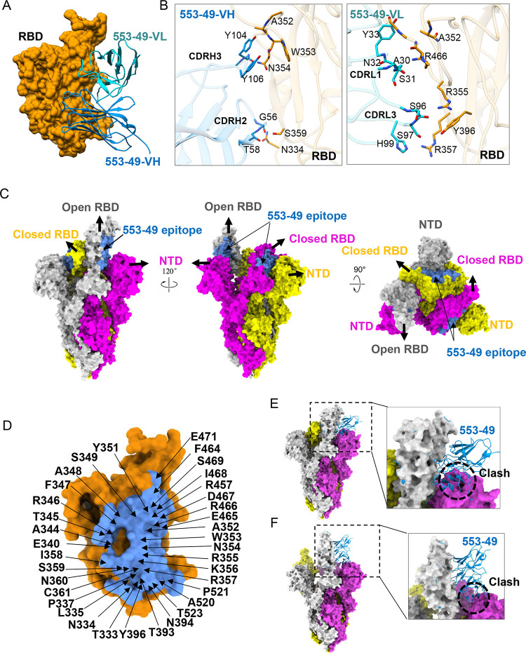
FIG 2.
Cryo-EM structure of SARS-CoV-2 Omicron S in complex with IgG 553-49. (A) Structure of Omicron S RBD–553-49. The RBD is displayed in orange surface mode. The heavy chain and light chain of 553-49 are shown as ribbons colored in cornflower blue and cyan, respectively. (B) The interfaces between the RBD and 553-49. (C) Molecular-surface representation of the apo-Omicron S model displayed in side and top views. The 553-49 epitope (colored in cornflower blue) on the RBD was covered by the NTD of the adjacent protomer. (D) Close-up view of the 553-49 epitope on the RBD. The residues involved in the interaction are labeled. (E, F) Binding of 553-49 to a spike trimer with one up RBD (E) or a wide-open RBD (PDB 7WHK) (F) would clash with the NTD of the adjacent protomer (indicated by the black dashed circles).

