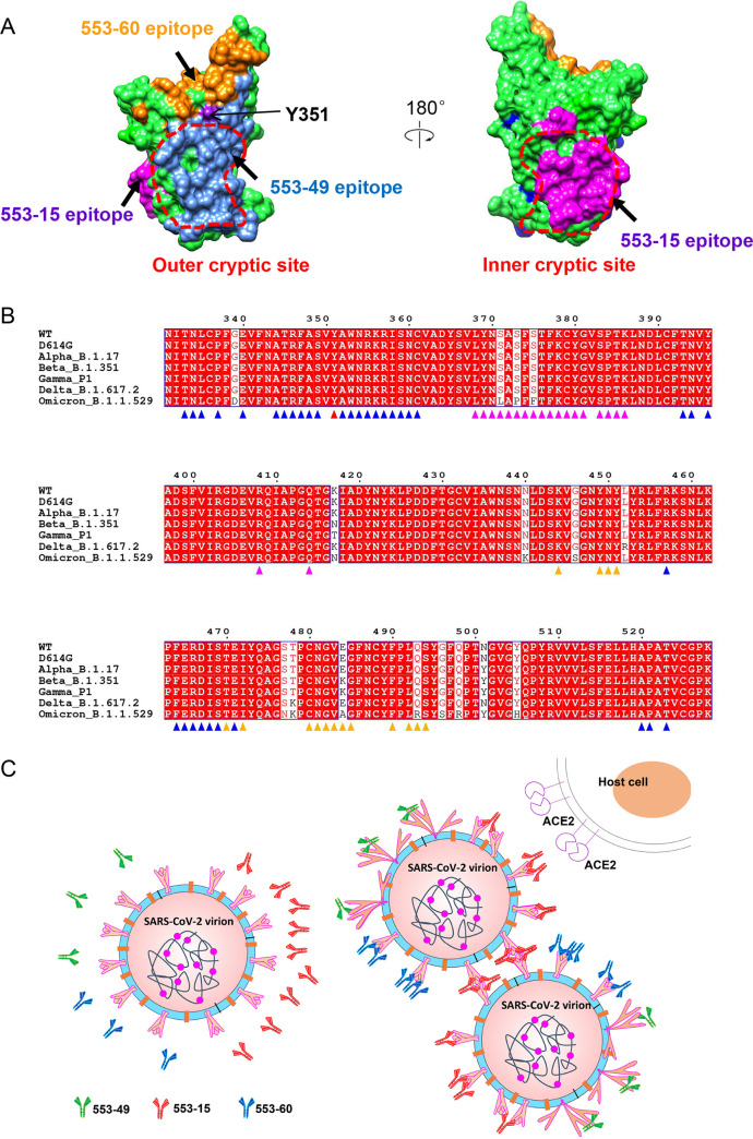
FIG 6.
Epitopes and neutralization mechanisms of three NAbs. (A) Comparison of 553-49, 553-15, and 553-60 epitopes on the S RBD. The RBD is displayed in green surface representation. The 553-49, 553-15, and 553-60 epitopes are colored cornflower blue, magenta, and orange, respectively. The inner and outer cryptic sites are indicated by red dotted lines. (B) Sequence alignment of S RBDs of SARS-CoV-2 WT and all VOCs, showing that IgG 553-49 targets to a completely conserved epitope. Conserved amino acids are highlighted in red. Residues involved in 553-49, 553-15, and 553-60 interactions are marked with triangles in blue, magenta, and orange, respectively. Residues involved in both 553-49 and 553-60 binding are marked with triangles in red. (C) Schematic model of the neutralization mechanisms of 553-49, 553-15, and 553-60.

