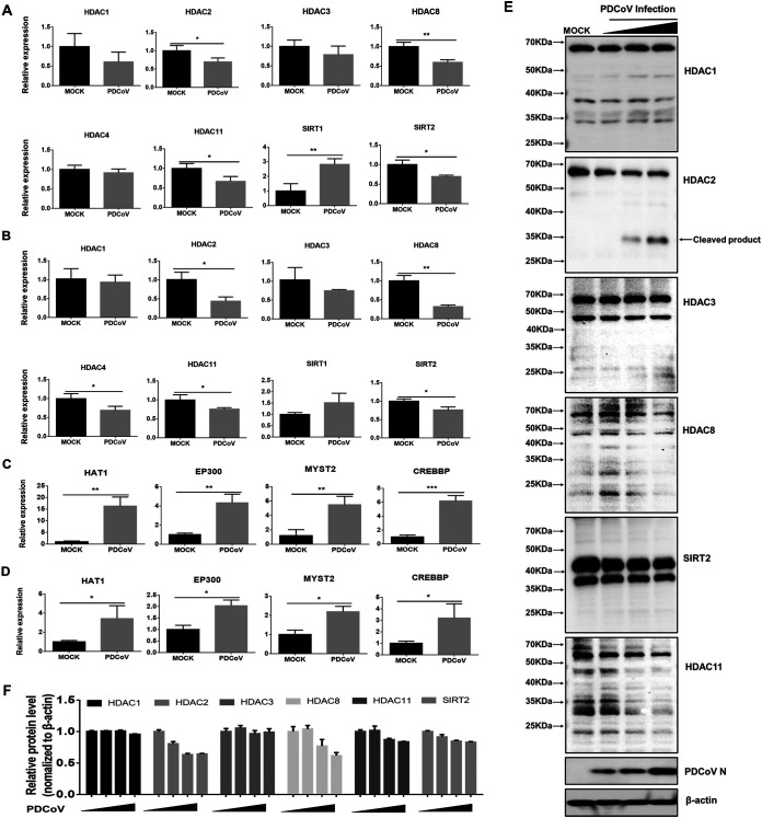
FIG 2.
PDCoV infection downregulates expression of some HDACs and upregulates expression of tested HATs. (A–D) LLC-PK1cells (A, C) and IPI-FX cells (B, D) were mock-infected or infected with PDCoV (MOI 1). At 24 hpi, the cells were collected for qRT-PCR to detect HDAC mRNA expression (A, B) and HATs (C, D). Relative mRNA expressions in PDCoV-infected cells were normalized to those of mock-infected cells. All experiments were performed in triplicate, and the data are presented as means ± SD of three independent experiments (*, P < 0.05; **, P < 0.01; ***, P < 0.001). (E) LLC-PK1 cells were infected with PDCoV at increasing infectious doses (MOI 0.25, 0.5, or 1). At 24 hpi, cells were collected for Western blotting with antibodies against HDAC1, HDAC2, HDAC3, HDAC8, SIRT2, and HDAC11. Anti-N protein antibody was used to confirm PDCoV infection. (F) Density analysis of panel E represents the relative HDACs expression levels compared to the control group. The analysis was performed with the ImageJ software package.

