FIG 4.
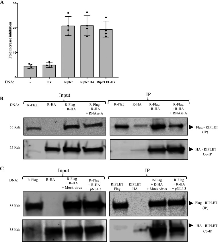
Riplet self-associates. (A) Fold increase of ZAP-mediated HIV-1 inhibition in cells expressing Riplet, Riplet-HA, and Riplet-Flag. 293TrexhZAP cells transfected with the indicated DNAs were infected with HIV-1 reporter virus followed by ZAP induction with doxycycline or DMSO as a control. Cells were lysed 4 h postinfection and assayed for luciferase. The fold virus inhibition was calculated as the ratio of luciferase expression levels in DMSO-treated cells to those in doxycycline-treated cells after normalization to total protein content by Bradford assay for each sample. Data points represent the mean ± SD values of four independent experiments. (B) Coimmunoprecipitation of tagged Riplet proteins. 293T cells were transfected with DNAs overexpressing Riplet-Flag (R-Flag) or Riplet-HA (R-HA) or both, as indicated, and lysed at 48 h posttransfection. Lysates were treated with RNase A (50 μg/mL). (B, Left) In the input data, total proteins were analyzed by gel electrophoresis, blotted, and probed for Flag-tagged Riplet with mouse α-Flag antibody (top) or HA-tagged Riplet with anti-HA antibodies (bottom). (B, Right) In the IP data, Flag-tagged Riplet was recovered by immunoprecipitation with α-Flag antibody, and bound proteins were analyzed by electrophoresis, blotted, and probed for Flag-tagged Riplet (top) or HA-tagged Riplet (bottom). Approximate molecular weights of major proteins estimated from size markers are indicated on left. (C) Coimmunoprecipitation of tagged Riplet proteins in 293TrexhZAP cells after various treatments. 293TrexhZAP cells were transfected with DNAs overexpressing Riplet-Flag (R-Flag) or Riplet-HA (R-HA) or both and infected with HIV-luc reporter virus (pNL4.3) or with a mock virus preparation lacking the VSVG envelope (mock virus). Lysates were prepared 48 h postinfection. (C, Left) In the input data, total proteins were analyzed by gel electrophoresis, blotted, and probed for Flag-tagged Riplet with mouse α-Flag antibody (top) or HA-tagged Riplet with anti-HA antibodies (bottom). (C, Right) In the IP data, Flag-tagged Riplet was recovered by immunoprecipitation with α-Flag antibody, and bound proteins were analyzed by electrophoresis, blotted, and probed for Flag-tagged Riplet (top) or HA-tagged Riplet (bottom). Approximate molecular weights of major proteins estimated from size markers are indicated on left.

