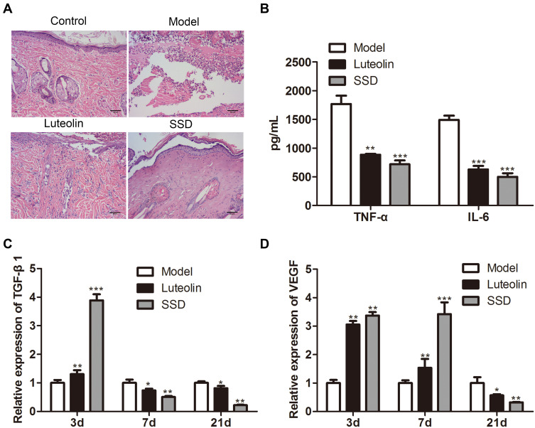
Figure 3.
The therapeutic effect of luteolin on scald model rats. (A) Hematoxylin-eosin staining used to analyze the histopathological changes of the scald wound. (B) The expression of TNF-α and IL-6 in serum evaluated by ELISA on day 21. The relative expression of (C) TGF-β1 and (D) VEGF in scald wound tissues detected by qRT-PCR on day 3, 7 and 21. The values were presented as mean ± SD (n=3). *P<0.05; **P<0.01 and ***P<0.001 versus the Model group.

