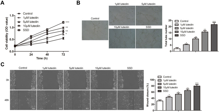
Figure 4.
The effect of luteolin on proliferation, angiogenesis and migration in HMEC-1. (A) HMEC-1 cells were treated with different concentration of luteolin (0, 1, 5, 10 μM) and SSD for 24, 48 and 72 h, the cell viability was analyzed by CCK-8 assay. (B) HMEC-1 were treated with different concentration of luteolin (0, 1, 5, 10 μM) and SSD for 8 h, tubule formation experiment was used to observe the lumen structure of HMEC-1 cells. (C) HMEC-1 cells were treated with different concentration of luteolin (0, 1, 5, 10 μM) and SSD for 48 h, migration was analyzed by scratch assays. Scale bar =200 μm. The values were presented as mean ± SD (n=3). *P<0.05; **P<0.01 and ***P<0.001 versus the control group.

