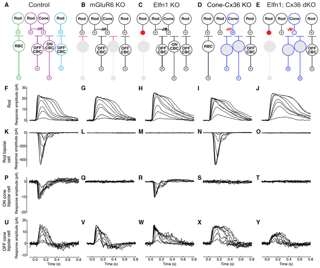
Figure 1. Rod and cone circuit pathways in control and mutant mice.

(A) A schematic of the three rod pathways in the mammalian outer retina. In the primary rod pathway (green), the rod signal is passed from rods to rod BCs (RBCs). In the secondary rod pathway (purple), the rod signal is passed from rods to cones via a gap junction connection and then from cones to ON and OFF cone BCs (CBCs). In the tertiary rod pathway (blue), the rod signal is passed from rods to OFF CBCs.
(B) A retinal circuit schematic of the mGluR6 knockout (KO) mouse showing functional and non-functional pathways. RBCs and ON CBCs cannot receive glutamate signals because of loss of metabotropic glutamate receptor 6 (mGluR6) at their dendrites (red). The downstream rod ON pathways no longer receive rod input (gray).
(C) A retinal circuit schematic of the Elfn1 KO mouse. Because of loss of the presynaptic protein Elfn1, the synapse between rods and RBCs is non-functional (red). The primary pathway no longer receives rod input (gray).
(D) A retinal circuit schematic of the Cone-Cx36 KO mouse. Because of loss of connexin 36 (Cx36) in cones, no gap junctions are formed between rods and cones (red). The secondary pathway no longer receives direct rod input (gray); indirect input from the primary rod pathway via the All amacrine cell (not pictured) is still functional (white synapses). Cone pathways, indicated by blue outlines, are functional.
(E) A retinal circuit schematic of the Elfn1; Cx36 double KO (dKO) mouse. The synapse between the rod and RBC is non-functional, and there is lack of rod-to-cone gap-junctional coupling (red). The primary and secondary rod pathways no longer receive rod input (gray). Cone pathways, indicated by blue outlines, are functional.
(F–J) Physiological recordings of rod photocurrents in retinal slices from control, mGluR6 KO, Elfn1 KO, Cone-Cx36 KO, and Elfn1; Cx36 dKO mice, respectively. Recordings were made in whole-cell patch-clamp mode (Vm = −40 mV). 20-ms light flashes were given at time 0 s. Flash strength ranged from 2.5–156 R*/rod. Recordings are representative of data collected across several cells (Table 1).
(K–O) Physiological recordings of light-evoked RBC responses. Recordings were made in the same slices as the rod recordings and as before, with some differences (Vm = −60 mV and flash strength from 0.2–16 R*/rod). RBC light responses were never observed in mGluR6 KO, Elfn1 KO, or Elfn1; Cx36 dKO mice (Table 1; Figure S3).
(P–T) ON CBC responses as described for RBCs. Flash strength spanned a range expected to activate the primary and secondary rod pathways. ON CBC light responses were never observed in mGluR6 KO, Cone-Cx36 KO, or Elfn1; Cx36 dKO mice (Table 1; Figure S3).
(U–Y) OFF CBCs responses as described for ON CBCs. Flash strength spanned a range expected to activate the primary, secondary, and tertiary rod pathways.
