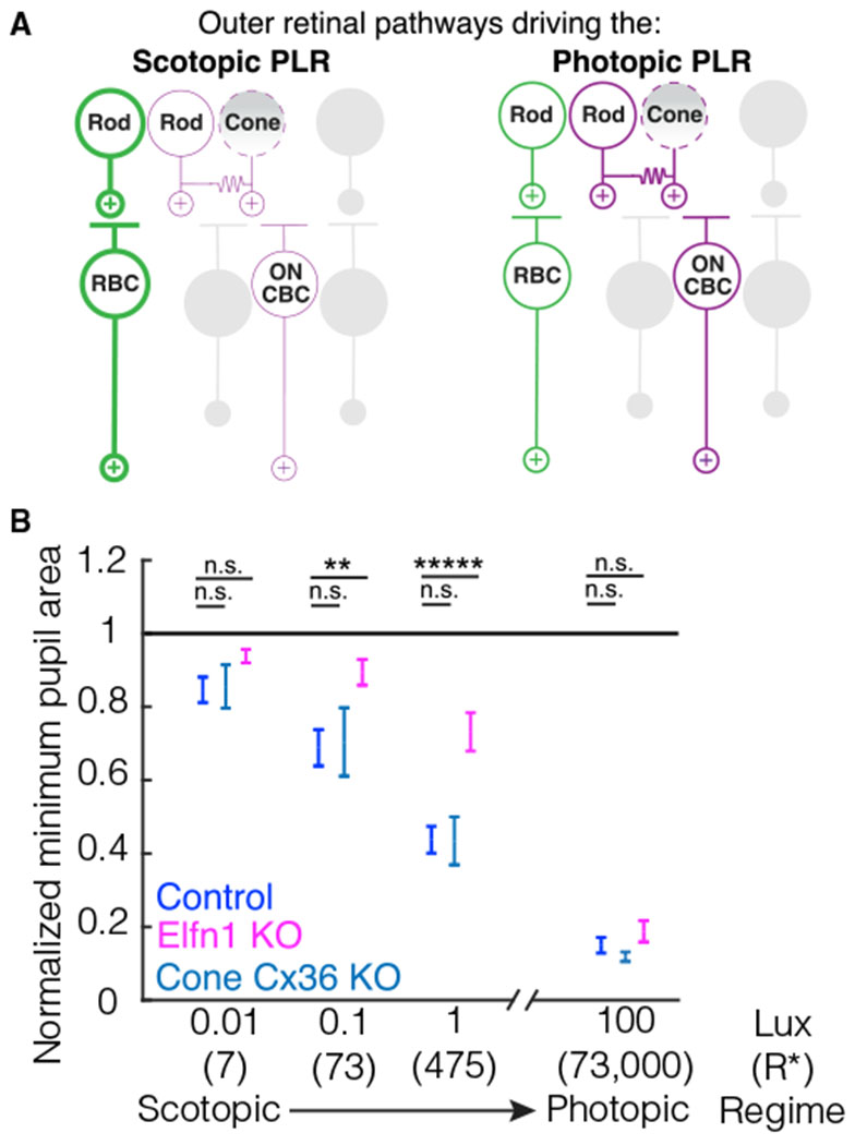
Figure 6. The outer retinal circuit pathways driving the PLR.

(A) A schematic detailing the outer retinal circuit pathways that drive the photopic and scotopic PLR. Left: the primary rod pathway is the predominant circuit driving the scotopic PLR (bold green) and is required for normal pupil constriction. The secondary ON rod pathway (purple) cannot completely compensate for the primary rod pathway. The secondary OFF rod pathway and tertiary rod pathway cannot drive the scotopic PLR (gray). Cones cannot compensate for rods at scotopic light levels (indicated by a dashed outline and shaded gradient). Right: the primary rod pathway (green) and secondary ON rod pathway (purple) can drive the normal photopic PLR. The secondary OFF rod pathway and tertiary pathway cannot drive the photopic PLR (gray). Cones cannot compensate for rods even when the secondary rod pathway drives the photopic PLR (indicated by a dashed outline and shaded gradient).
(B) Summary plot of control and mutant mouse pupil constriction across scotopic to photopic light levels. Data shown are summarized from Figures 3 and 5. Elfn1 KO mice, but not Cone-Cx36 KO mice, have deficits in their PLR under scotopic and mesopic light regimens. Error bars indicate SEM. Significance from the control group at each light level is as follows (ANOVA post hoc Dunnett’s method): at 100-lux, Elfn1 KO p = 0.58, Cone-Cx36 KO p = 0.89; at 1-lux, Elfn1 KO p = 1.3 × 10⁻⁴ Cone-Cx36 KO p = 1; at 0.1-lux, Elfn1 KO p = 0.008, Cone-Cx36 KO p = 0.98; at 0.01-lux, Elfn1 KO p = 0.33, Cone-Cx36 KO p = 0.99.
