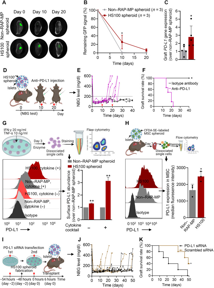
Fig. 7. The essential role played by MSC-mediated PD-L1 on islet xenograft survival.
(A and B) Retention of GFP-expressing MSCs in islet xenografts over time (values are residual GFP intensities; n = 3). (C) Relative gene expression of PD-L1 in whole grafts containing islets and non–RAP-MP or HS100 spheroids at 12 days after transplantation (n = 5). (D to F) The effect of anti–PD-L1 antibody treatment (2.5 mg/kg per dose × 2 doses, intraperitoneally delivered on days 10 and 20 after transplantation; n = 6) on mice cotransplanted with islets and HS100 spheroids. (D) Graphical illustration of the experimental design, (E) NBG test results, and (F) Kaplan-Meier curves for islet xenograft survival times. Transplanted mice treated with isotype control antibody served as controls (n = 3). (G and H) In vitro and in vivo PD-L1 protein expressions on MSCs in non–RAP-MP and HS100 spheroids, as determined by flow cytometry. (G) Spheroids were treated with or without a cytokine cocktail for 3 days before the assessment (n = 2 per group). (H) MSCs were labeled with CFDA-SE before fabricating spheroids. Grafts were retrieved at 7 days after transplantation (n = 3). MSCs were considered to express PD-L1 if double-positive for CFDA-SE and PD-L1. (I to K) Effect of MSC-mediated PD-L1 on islet xenograft survival. (I) Graphical illustration of the experimental design. MSCs were transfected twice with 50 nM PD-L1 siRNA or 50 nM scrambled siRNA before transplantation, (J) NBG test results, and (K) Kaplan-Meier curves of islet xenograft survival times (n = 5). Data are expressed as the means ± SDs. Data in (B) were analyzed using two-way ANOVA, in (C), (G), and (H) using the unpaired two-tailed t test, and in (F) and (K) using the log-rank (Mantel-Cox) test.

