. 2022 Aug 1;11:e80130. doi: 10.7554/eLife.80130

Figure 3. Force-dependent binding interactions between the αE-catenin actin-binding domain (ABD) and F-actin.

(A) (Top) GFP-haloligand and fusion protein Halotag-ABD (red) complexes are immobilized on silica microspheres attached to a microscope coverslip. A taut actin filament is suspended between two optically trapped beads and held over the assembled complexes. The stage is translated parallel to the actin filament, and when at least one protein complex binds to F-actin, the trapped beads are pulled out of their equilibrium position. The restoring force of the optical trap (black arrows) applies tension on a bound complex while bystander complexes (pale) bind and unbind transiently. (B) A representative force versus time series for the constant-force assay. (Top) Plotted are the forces summed from both traps versus time, decimated from 40 to 4 kHz. We observe traces characterized either by rupture of a single bound molecule (left) or by sequential rupture of multiple bound molecules (right). Traces colored in black are regions used for force baseline determination, and vertical lines indicate step boundaries. (Bottom) If summed forces surpass a threshold, stage motion halts until detachment of the final bound molecule. (C) αE-catenin ABD forms a catch bond with F-actin (N = 900). Areas of all circles are proportional to the number of events measured in each equal-width bin. These data are represented here without depicting the direction of force applied relative to the polar actin filament.

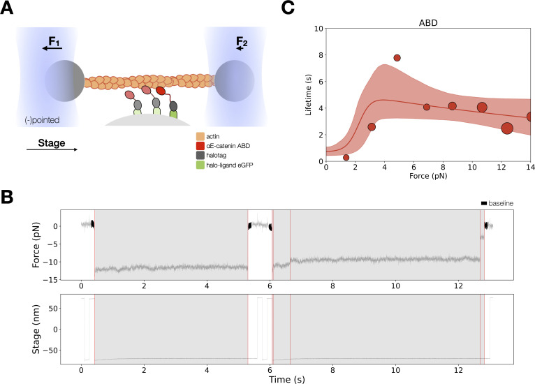
Figure 3—source data 1. αE-catenin actin-binding domain (ABD) optical trap constant-force assay multi-step force versus lifetimes.

Figure 3.

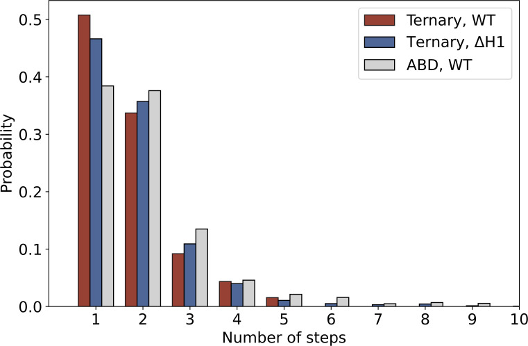
Figure 3—figure supplement 1. The distribution of the number of steps in constant-force assay measurements of ternary wild type (red, N = 1418), ternaryΔH1 (blue, N = 1604), and actin-binding domain (ABD) (gray, N = 1460).

Figure 3—figure supplement 1.

The probability of observing single- versus multi-step events is comparable between ternary wild type and ternaryΔH1.
Figure 3—figure supplement 1—source code 1. Step number distribution analysis code for all events.
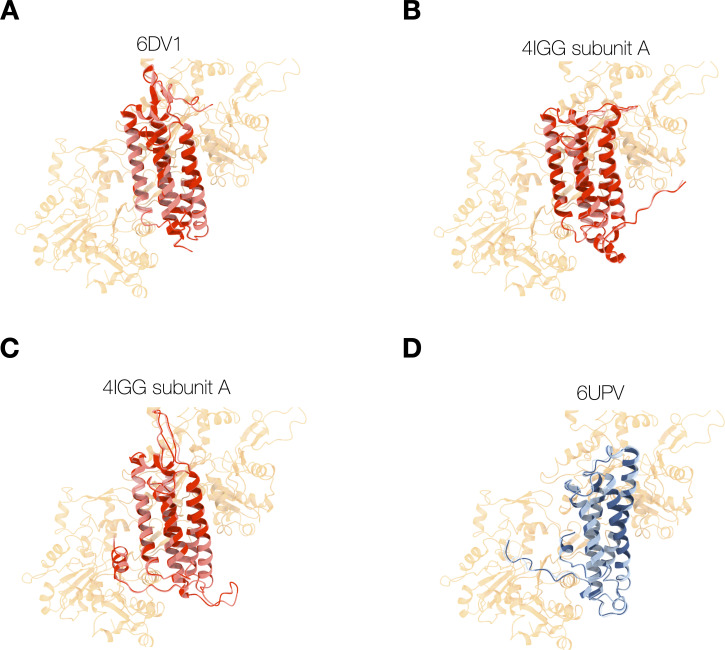
Figure 3—figure supplement 2. Energy-minimized actin-binding domain (ABD) structures superimposed (red) or bound (blue) with actin.

Figure 3—figure supplement 2.

(A–C) Structures of isolated ABD (PDB entries 6dv1, 4igg chain A, 4igg chain B) were superimposed on the actin-bound ABD (PDB entry 6UPV), and minimized while docked to actin (red), or in the absence of actin (pink). Energy minimization of these ABDs bound to actin resulted in minor structural changes (RMSD <0.7 Å) and resolved clashes with overlapping atoms. (D) Structures of the actin-bound ABD (PDB entry 6UPV) minimized in the presence (navy) and absence (light blue) of actin.
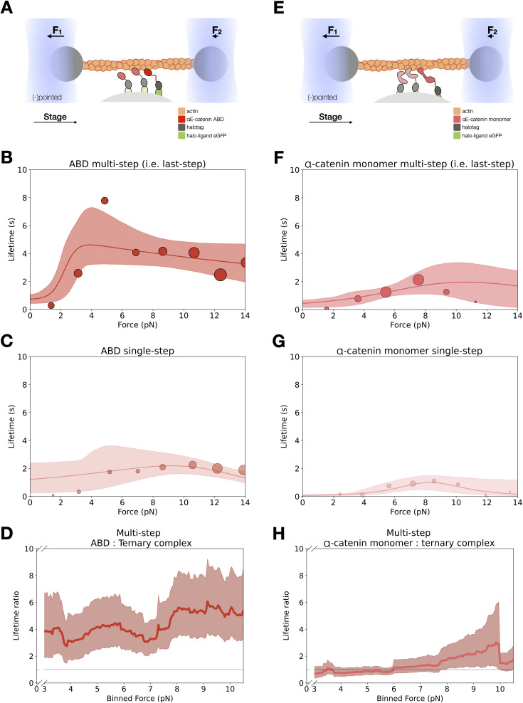
Figure 3—figure supplement 3. Force-dependent binding lifetimes of αE-catenin actin-binding domain (ABD) and monomer.

Figure 3—figure supplement 3.

(A) GFP-haloligand and fusion protein Halotag-ABD (red) complexes are immobilized on silica microspheres and interact with a taut actin filament. This figure is the same as Figure 3A. (B) Force-dependent binding lifetimes of αE-catenin ABD to F-actin, for the last detachment step in multi-step events. This figure is the same Figure 3C. (C) Force-dependent binding lifetimes of αE-catenin ABD to F-actin in single-step events. (D) Computed lifetime ratios (LRs) between ABD and the ternary complex with a 4 pN sliding window across 0–13 pN. The ABD:F-actin-binding interaction is fourfold longer than that of the ternary complex (mean LR = 4.27, 90% confidence interval [CI] = 2.55–6.67). (E) GFP-haloligand and fusion protein Halotag-αE-catenin (pink) complexes are immobilized on silica microspheres and interact with a taut actin filament. (F) Force-dependent binding lifetimes of αE-catenin monomer to F-actin, for the last detachment step in multi-step events (N = 442). (G) Force-dependent binding lifetimes of αE-catenin monomer to F-actin in single-step events (N = 140). (H) Computed LRs between αE-catenin monomer and the ternary complex with a 4 pN sliding window across 0–13 pN. The αE-catenin:F-actin-binding interaction is comparable to the ternary complex (mean LR = 1.34, 90% CI = 0.77–2.20).
Figure 3—figure supplement 3—source data 1. αE-catenin actin-binding domain (ABD) optical trap constant-force assay single-step force versus lifetimes.
Figure 3—figure supplement 3—source data 2. αE-catenin monomer optical trap constant-force assay single-step force versus lifetimes.
Figure 3—figure supplement 3—source data 3. αE-catenin monomer optical trap constant-force assay multi-step force versus lifetimes.
Figure 3—figure supplement 3—source data 4. Bootstrapped lifetime ratios for actin-binding domain (ABD) versus ternary complex multi-step data.
Figure 3—figure supplement 3—source data 5. Bootstrapped lifetime ratios for αE-catenin monomer versus ternary complex multi-step data.