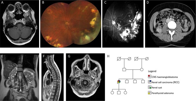
Figure 1.
(A) Axial T1-weighted post-contrast image through the orbits shows a retinal angioma in the left globe. (B) Colour fundus photograph of left eye at most recent clinic visit showing areas of previous laser and cryotherapy treatment with dragging of optic nerve vessels towards inferotemporal quadrant. Haemangiomas present in macula and nasal quadrant. Multiple peripheral chorioretinal scars related to previously treated haemangiomas. Superotemporal vessels with perivascular exudate. Right eye normal (not shown). Visual acuity Right 6/5 and Left 6/12. (C) Fluorescein angiogram performed 4 years previously showing areas of scarring and retinal detachment related to exudation and effect of treatment. Optic nerve leak related to the effect of traction and glial proliferation with areas of hyperfluorescence in the macula related to new small haemangiomas, visible anterior to the internal limiting membrane on OCT scans (not shown). Right eye normal (not shown). (D) CT scan showing 29 mm diameter RCC in right kidney. (E) Coronal T2-weighted image through the abdomen shows numerous small cysts in both kidneys. (F) Sagittal T1-weighted post-contrast image of the spinal cord shows a solid enhancing haemangioblastoma with associated hypertrophied vessels on the dorsal surface of the spinal cord. (G) Axial T1-weighted post-contrast image through the cervicomedullary junction shows a small solid haemangioblastoma. (H) Family pedigree of sporadic case of VHL.

