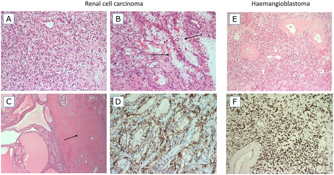
Figure 2.
Haematoxylin and eosin (H&E)-stained images (A–C) and CA-IX staining image from the RCC. H&E and CD34-stained images from the haemangioblastoma. (A) An area with typical features of a ccRCC, composed of a sheet of small cells with clear cytoplasm and a delicate background vascular network. (B) Focus on branching tubules in which the tumour cells have more voluminous clear cytoplasm (arrows). (C) A cystic area of the RCC tumour (left) and a dense band of leiomyomatous (muscular) stroma (right, arrow). (D) The tumour was diffusely positive for CA-IX, a classic marker of HIF up-regulation. (E) The haemangioblastoma tumour is composed of very small cells with clear cytoplasm and a background vascular network. Larger blood vessels have thickened hyalinized walls. (F) The vascular network of haemangioblastoma is highlighted by CD34 immunohistochemistry.

