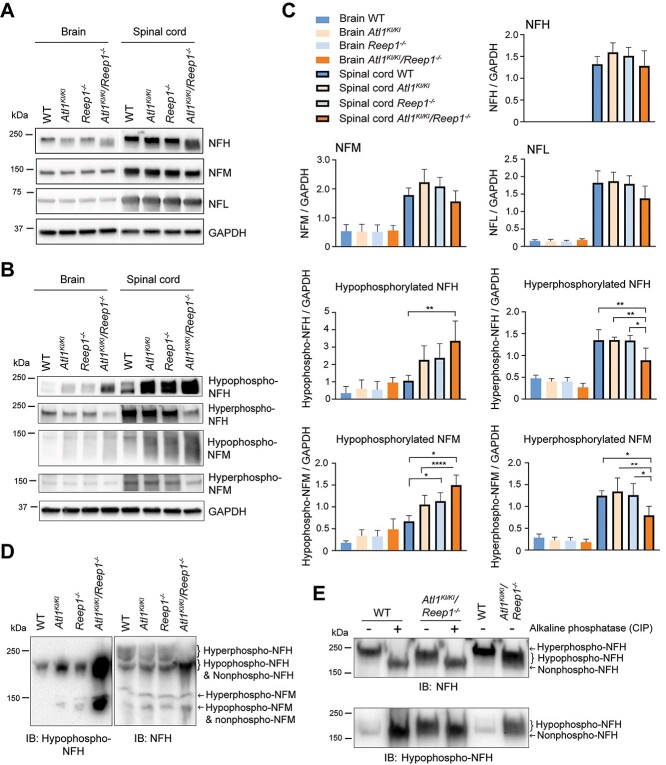
Figure 7.
Hypophosphorylation of neurofilaments in Atl1KI/KI/Reep1−/− mice. (A, B) Lysates of brain and spinal cord from 6-month-old WT mice and the indicated mutant genotypes were immunoblotted as shown for total neurofilaments (A) and those with different phosphorylation states (B). GAPDH levels were monitored as a control for protein loading. (C) Quantifications of immunoblots from A and B. One-way ANOVA (Tukey’s test) was applied for statistical analyses (n = 4). *P < 0.05, **P < 0.01, ****P < 0.001. (D, E) Changes of phosphorylation levels were further analyzed using Phos-tag SDS-PAGE (D) and by adding CIP to sample lysates to dephosphorylate neurofilaments (E).

