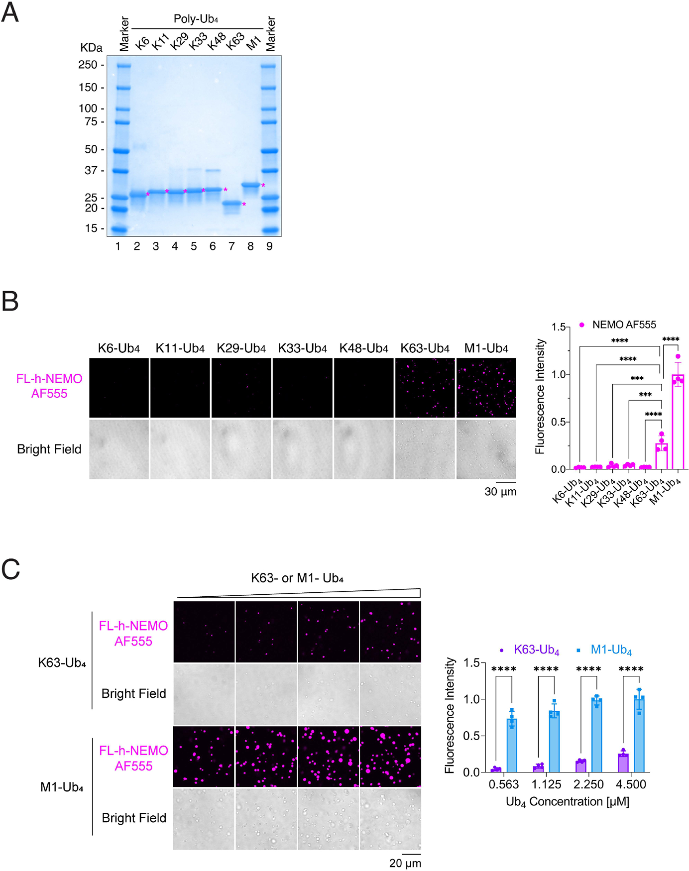
Figure 2. NEMO phase separation is specifically induced by K63-linked and linear polyubiquitin chains.

(A) Coomassie blue staining of Ub4 linked through K6, K11, K29, K33, K48, K63 or M1. Asterisks indicate the protein of interest (Ub4).
(B) Left panel: representative fluorescent images of NEMO liquid droplets induced by Ub4 linked through K6, K11, K29, K33, K48, K63, and M1. Liquid droplets formed after mixing of 6 μM human FL-NEMO (3% was labeled with Alexa 555) with 4.5 μM Ub4 of different linkages for 30 minutes at 37°C. Right panel: quantification of fluorescence intensity of liquid droplets.
(C) Left panel: representative fluorescent images of NEMO liquid droplets induced by K63- or M1- Ub4 at different concentrations. Liquid droplets formed after mixing of 6 μM human FL-NEMO (3% was labeled with Alexa 555) with indicated concentrations of K63- or M1- Ub4 for 45 minutes at 37°C. Right panel: quantification of fluorescence intensity of liquid droplets.
Data shown in (B) and (C) are means ± SD. n = 4 areas. In (B), one-way analysis of variance (ANOVA); in (C), two-way analysis of variance (ANOVA). ***P < 0.0002; ****P < 0.0001.
See also Figure S3.
