
Extended Data Figure 4. NLRP3 activity is strongly associated with dTGN but not mitochondria.
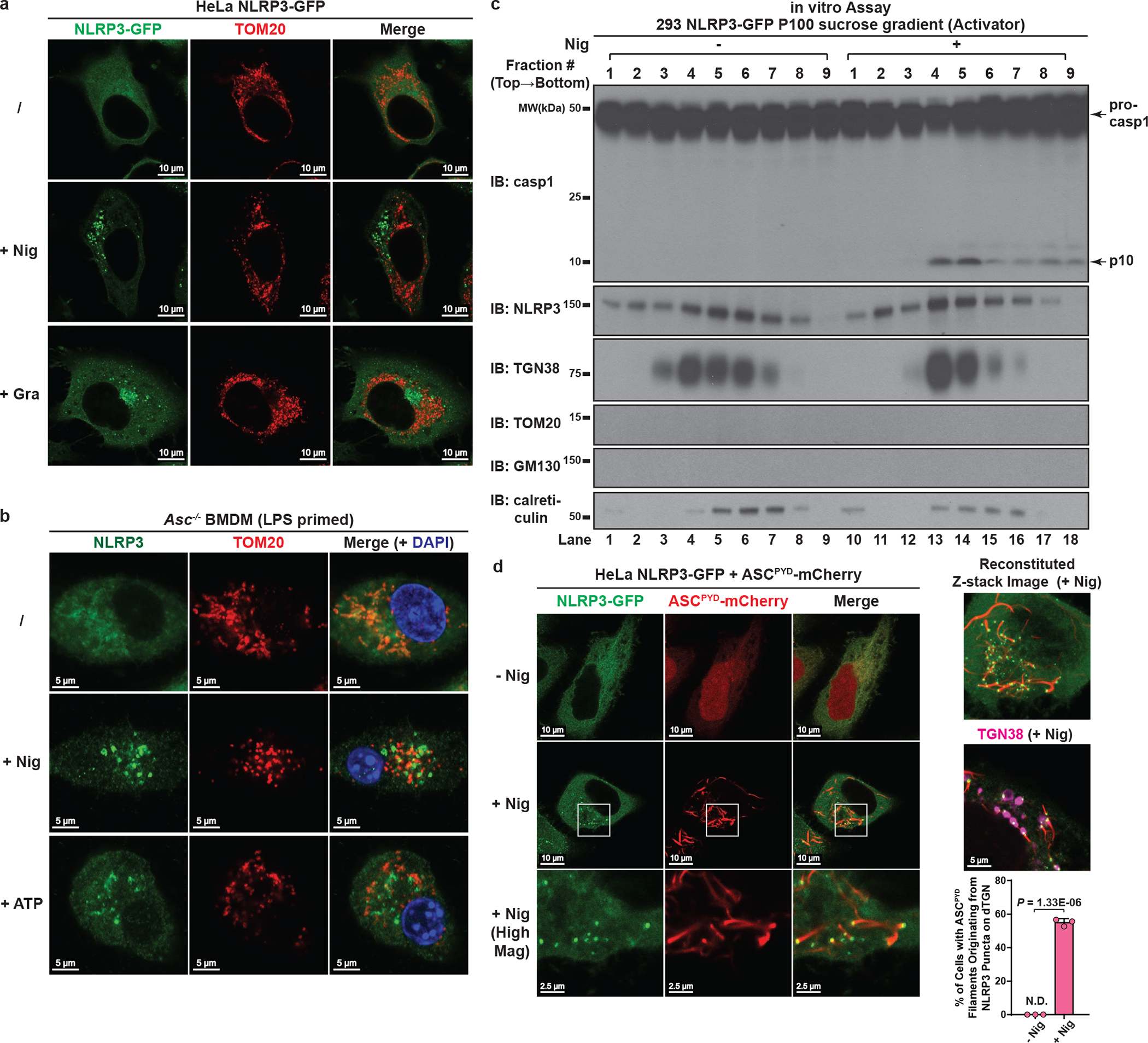
a, NLRP3 did not translocate to mitochondria upon stimulation in HeLa cells. HeLa NLRP3-GFP cells were stimulated with nigericin (10 μM) or gramicidin (5 μM) for 80 min before immunostained for TOM20 (mitochondrial marker). b, Neither nigericin- nor ATP-induced NLRP3 puncta were colocalized with mitochondria in ASC-deficient BMDMs. Cells were primed with LPS (50 ng/mL) for 3 hours, followed by nigericin (10 μM) or ATP (5 mM) treatment for 60 min before immunostained for endogenous NLRP3 and TOM20. c, NLRP3 activity in P100 (light membrane) fraction was strongly associated with dTGN but not mitochondria. P100 fraction collected from Fig. 1c was subjected to sucrose gradient ultracentrifugation, before fractions were collected and examined by the in vitro NLRP3 activity assay (top panel). TOM20 (mitochondrial marker) and GM130 (cis-Golgi marker) were not detectable on immunoblots even after prolonged exposure. d, dTGN-localized NLRP3 puncta can initiate aggregation of ASCPYD. HeLa cells stably expressing the indicated proteins were incubated −/+ nigericin (10 μM) for 80 min before imaging. Mag, magnification. ASCPYD: aa 1–90 of murine ASC. Right panel (from top to bottom): reconstituted Z-stack image of a representative nigericin-treated cell; nigericin-treated cell co-immunostained for TGN38 (pseudo-colored in magenta); percentage of cells with ASCPYD filaments originating from dTGN-localized NLRP3 puncta was quantified from 100 cells (n = 3, mean ± SD, two-sided t test). N.D., not detectable.