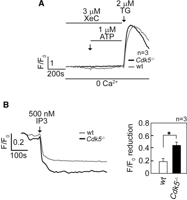
Fig. 2.

Increased [Ca2+]cyt in Cdk5−/− MEFs is due to increased IP3R-mediated Ca2+ release. A MEFs loaded with Fluo-4 AM and treated with 3 µM XeC followed by 1 µM ATP then 2 µM TG in Ca2+-free EGTA-containing KRH buffer were analyzed for [Ca2+]cyt transients by single-cell Ca2+ imaging analyses. The similar increase in [Ca2+]cyt in wt and Cdk5−/− MEFs upon treatment with TG, which was added after 15 min of treatment with XeC, indicates comparable viability of these cells during analysis. Data represent means of Ca2+ signal traces from 15 cells. B Measurement of Ca2+ release from internal stores upon IP3 treatment is described in Materials and methods. Ca2+ release was measured every 4 s by single-cell Ca2+ imaging. Data represent means of Ca2+ signal traces from ten cells, and are results from one of three independent experiments showing similar patterns. Values are means ± SEM from the three separate experiments (n = 3). *p < 0.05
