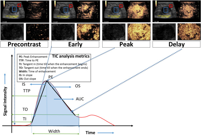
Fig. 1.
Top panel: CEUS scans showing 4 enhancement phases (precontrast, early, peak and delay) of the Invasive Ductal Carcinoma (ER-/PR-/HER2-) in a 44 year-old woman presenting with a left breast large palpable lump measuring up to 4.4 cm. The CEUS scan shows avidly enhancing irregular mass with marked heterogeneous enhancement and small areas of clear defect, which is worrisome for a malignant mass. On the CEUS clip, the breast mass (blue contour) and adjacent normal tissue (red contour) are annotated by a fellowship trained radiologist using ImageJ software. Middle panel: Segmented regions of interest (here, the lesion) from the CEUS scans are shown to highlight the variation in enhancement across the different phases. Bottom panel: Based on a normalized map of the breast mass, obtained by dividing the breast mass (blue contour in top panel) values by mean of the normal tissue (red contour in top panel) data, a surrogate mean time-intensity curve was acquired for the whole cine clip. Subsequently, representative images corresponding to four time points on the surrogate mean time-intensity curve were extracted from cine clips: precontrast, early, peak, and delay enhancement, respectively. Here, the four shortlisted normalized images show nodular enhancement of the breast mass (blue contour in top panel)

