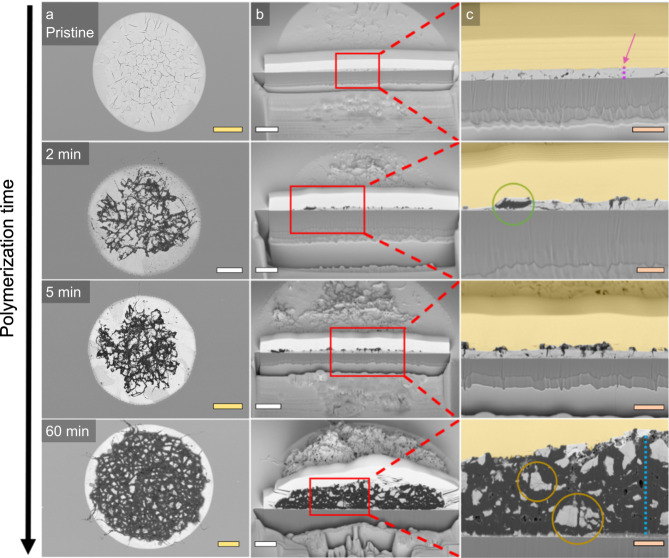
Fig. 4. Cross-sectional scanning electron microscopy (SEM) to study the fragmentation behaviour of the ethylene polymerized LaOCl spherical caps.
a Top-down overview of the full spherical caps at respectively 0 (pristine), 2, 5 and 60 min of ethylene polymerization. b A cross-section of the spherical caps that shows the internal structure of the polyethylene-LaOCl spherical cap composites. c A zoom-in of these cross-sections to provide enhanced view of the internal structure. The pink and blue dashed lines in respectively the pristine and 60 min ethylene polymerized samples show a thickness of 600 nm for the LaOCl spherical cap and 5.7 microns for the polyethylene layer at those positions. The green circle refers to LaOCl fragments having been lifted up from the spherical cap by the polyethylene formed within the porous catalyst framework. The orange circles indicate the internal cleavage sites of the LaOCl framework. The yellow scale bars represent 10 µm, the white scale bars 5 µm and the orange scale bars 2 µm. A yellow, transparent overlay is provided that indicates the coated Pt layer, all though in some cases it is observed to penetrate into the porous polyethylene layer.

