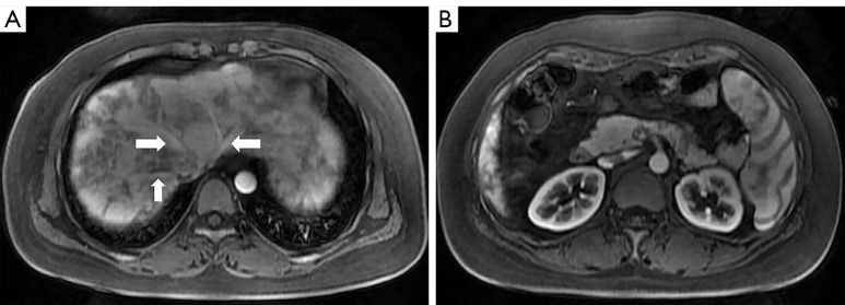
Figure 5.
The arterial phase of Gd-EOB-DTPA enhanced MRI. (A) Axial contrast-enhanced T1-weighted imaging showed hepatic venous trunks (arrows) were early enhanced, which were supposed to be enhanced in the portal phase. The liver enhancement was heterogeneous with a mosaic pattern of perfusion. (B) Axial contrast-enhanced T1-weighted imaging at a lower level than (A). The enhancement pattern of the kidneys and spleen confirmed that this enhancement phase was the arterial phase. Gd-EOB-DTPA, gadolinium-ethoxybenzyl-diethylenetriamine pentaacetic acid; MRI, magnetic resonance imaging.

