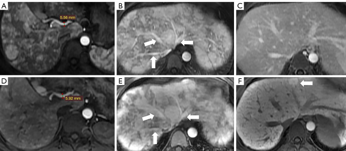
Figure 7.
The comparison of imaging features of liver involvement in HHT between the ECA-MRI and HBA-MRI. The imaging features of dilated and tortuous hepatic arterial branches (A,D), early filling of portal or hepatic venous trunks (B,E, arrows), and heterogeneous enhancement of parenchyma in the arterial phase (B,E) had no difference in two different contrast agents MRIs. The hepatic nodule showed high signal intensity (F) in the liver-specific phase of HBA-MRI, which was shown more clearly than in the delayed phase of ECA-MRI (C). (A) The early arterial phase of ECA-MRI; (B) the late arterial phase of ECA-MRI; (C) the delayed phase of ECA-MRI; (D) the early arterial phase of HBA-MRI; (E) the late arterial phase of HBA-MRI; (F) the liver-specific phase of HBA-MRI. HHT, hemorrhagic telangiectasia, MRI, magnetic resonance imaging; ECA-MRI, extracellular contrast agent MRI; HBA-MRI, hepatobiliary contrast agent MRI.

