
Figure 3. Lung-infiltrating monocytes acquire a tissue-resident phenotype and contribute to the increased AM population after N. brasiliensis inoculation.
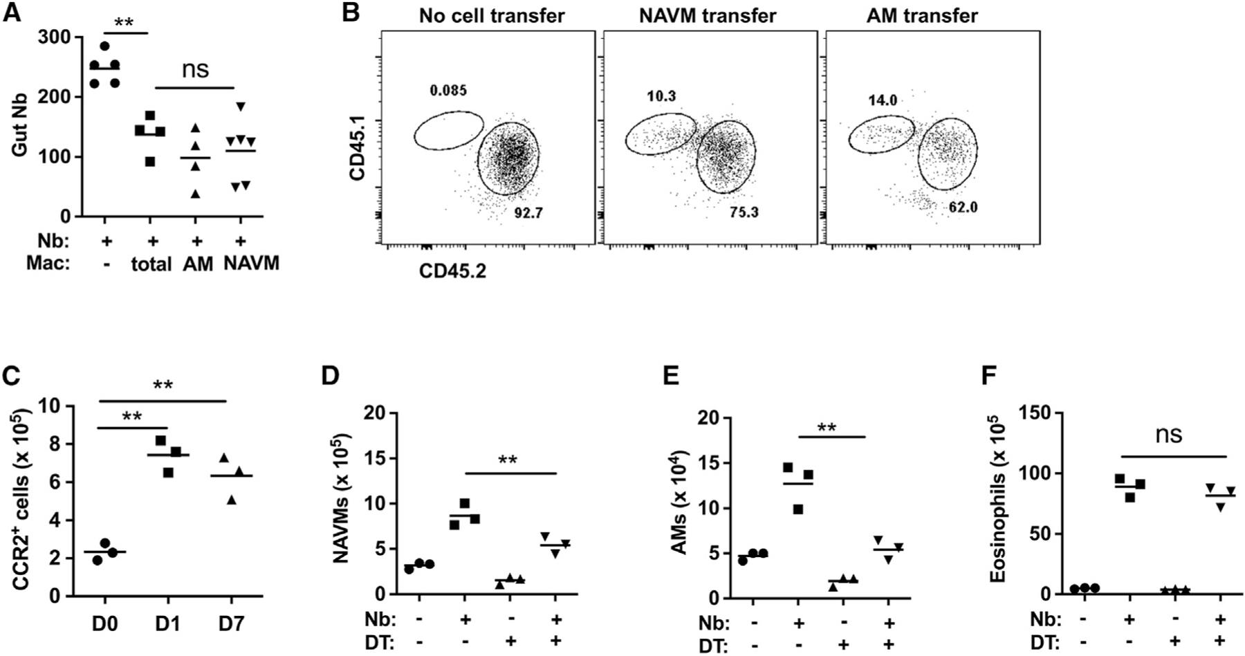
(A) At day 7 after N. brasiliensis (Nb) inoculation, donor macrophages including total macrophages, AMs, and NAVMs were electronically sorted and transferred to recipient mice, which were inoculated with Nb 2 days later. Recipient mice were assayed for intestinal worm numbers at day 5 after inoculation; a control group did not receive macrophages.
(B) Donor CD45.1 mice were inoculated with Nb, and 7 days later, lung NAVMs and AMs (as described in Figure 1) were sort-purified and transferred intratracheally (i.t.) into naive recipients (CD45.2), which were inoculated with Nb 2 days later. Donor and recipient lung AM populations were assessed at day 5 after Nb inoculation. Flow cytometry plots were representative of 3 mice per group and 2 independent experiments.
(C) CCR2-GFP reporter mice were assessed by flow cytometric analysis for monocyte recruitment to total lungs at days 1 and 7 after Nb inoculation.
(D–F) Monocytes were depleted by administration of DT at −1, +1, and +3 after Nb inoculation of CCR2-DTR mice. At day 7 after Nb inoculation, numbers of total lung NAVMs (D), AMs (E), and eosinophils (F) were determined by flow cytometric analysis.
Each symbol represents an individual mouse, and horizontal lines indicate the mean. All results are representative of two independent experiments. **p < 0.01 (one-way ANOVA).