Figure 3.
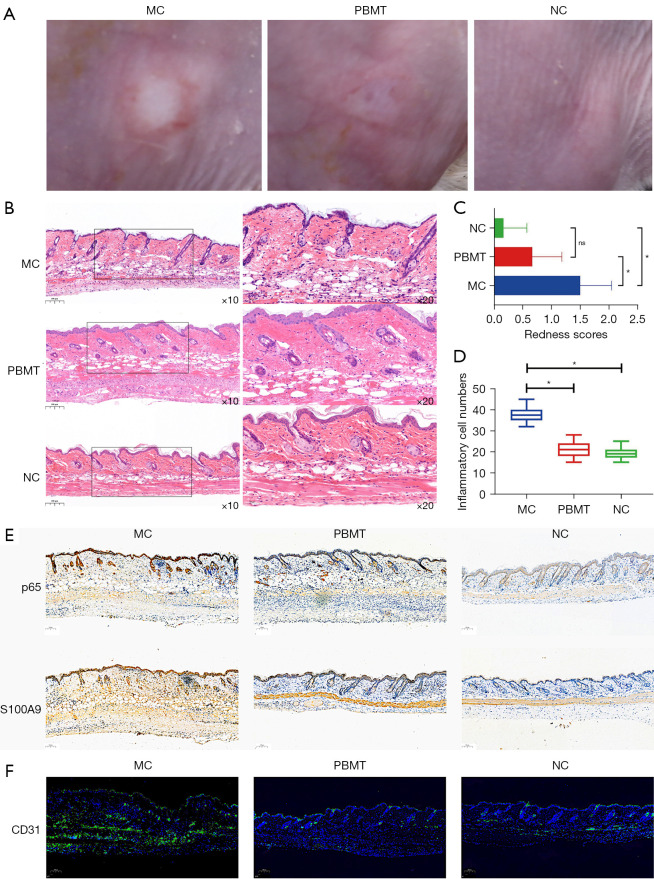
PBMT attenuated the severity of erythema, the inflammatory response, and angiogenesis in rosacea-like mouse skin in BALB/c mice. (A) Photographs of the dorsal skin lesions from the MC, PBMT, and NC groups. (B) H&E staining of skin lesions from the MC, PBMT, and NC groups. Scale bar =100 µm. (C) The redness scores of the skin lesions in the MC, PBMT, and NC groups (n=18, *, P<0.05; ns, not significant). (D) The inflammatory cell counts of the MC, PBMT, and NC groups based on H&E staining (n=18, *, P<0.05). (E) The expression of p65 and S100A9 in skin lesions from the MC, PBMT, and NC groups by immunohistochemistry. Scale bar =100 µm. (F) The expression of CD31 in the skin tissue from the MC, PBMT, and NC groups by immunofluorescence staining. Scale bar =100 µm. MC, model control; NC, negative control; PBMT, photobiomodulation therapy; H&E, hematoxylin and eosin.

