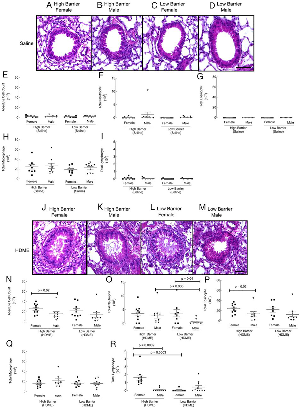
FIGURE 1. Airway histology and BAL cell counts.

Paraffin-embedded lung tissue sections were stained with H&E (A–D control groups; J–M HDME groups). Scale bar, 50 μm. Airways were lavaged with 0.7 ml of 2% FBS in saline. BAL cytospins were quantified for absolute cell numbers (E and N), neutrophils (F and O), eosinophils (G and P), macrophages (H and Q), and lymphocytes (I and R). Each dot represents data from one mouse. Ten mice in each group were used.
