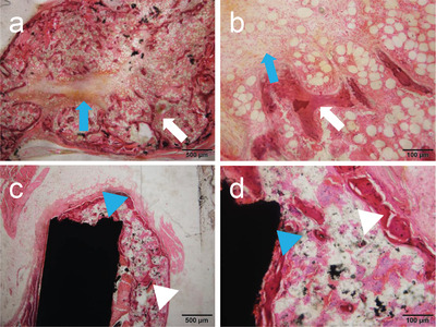
Figure 4.

Histological image at 3 weeks after implantation of rhBMP‐2‐containing MWCNT blocks in mouse back muscle. a,b) Ectopic bone formed around an rhBMP‐2‐containing PET‐fiber‐reinforced collagen sheet. Blue arrow: remnant of the PET‐fiber‐reinforced collagen sheet. White arrow: trabecular structure. c,d) Ectopic bone formed around an rhBMP‐2‐containing MWCNT block. The MWCNT block and newly formed bone are bound together firmly. Although the formed bone is found to contain a small amount of MWCNTs, no MWCNTs are detected in the surrounding tissue along with no inflammatory reactions. Blue arrowhead: MWCNT block. White arrowhead: trabecular structure. Hematoxylin‐eosin staining. Reproduced under the terms of the Creative Commons CC‐BY license.[ 86 ] Copyright 2017, The Authors. Published by PLOS.
