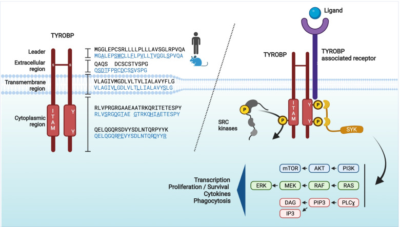
Fig. 1.
TYROBP structure and signaling pathway. Left panel: Mouse and human TYROBP: the TYROBP protein consists of a 27-aa leader peptide, a small 14-aa (16-aa in mouse, in blue) extracellular region, a 24-aa transmembrane segment, and a 48-aa (49-aa in mouse) cytoplasmic domain. Because of two cysteine residues (Cys33/35) in its extracellular region, TYROBP forms a disulfide-bonded homodimeric complex. TYROBP contains an immunoreceptor tyrosine-based activation motif (ITAM) in its cytoplasmic region. Right panel: Following ligand-binding by a TYROBP-associated receptor, the ITAM motif can be phosphorylated on its two conserved tyrosine residues by SRC kinases and induce the intracellular recruitment and activation of the spleen tyrosine kinase (SYK). Upon SYK recruitment, several downstream effector molecules such as phosphatidylinositol 3-kinase (PI3K), the small GTPase RAS, or the phospholipase Cγ (PLCγ) are mobilized, resulting in activation of transcription, proliferation, release of cytokines, and phagocytosis. Abbreviations: DAG, diacylglycerol; ERK, extracellular signal-regulated kinase; IP3, inositol trisphosphate; ITAM, immunoreceptor tyrosine-based activation motif; MEK, mitogen-activated protein kinase kinase; PI3K, phosphatidylinositol 3-kinase; PIP3, phosphatidylinositol-3,4,5-trisphosphate; PLCγ, phospholipase Cγ; SYK, spleen tyrosine kinase; TYROBP, tyrosine kinase binding protein; Y, Tyrosine

