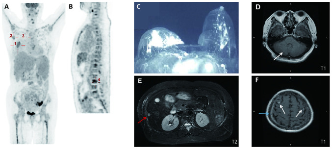
Figure 1.
Baseline imaging presentation. (A) PET/CT (10 days after initial presentation; coronal section) indicated a malignant tumor in the right breast (30.1×25.3 mm2; SUVmax, 5.2; marked by cross 1) with right-sided axillary (20.7×11.2 mm2; SUVmax, 3.2; and 17.4×9.8 mm; SUVmax, 6.0; marked by cross 2) and internal mammary (19.1×11.5 mm2; SUVmax, 2.6; marked by cross 3) metastatic lymph nodes. (B) PET/CT (median sagittal section) indicated systemic bone metastases in spine (marked by cross 4), bilateral ribs and sternum. (C) Contrast-enhanced breast CT (at the initial presentation) indicated an irregular mass with an unclear margin in the areola area of the right breast (31×15×22 mm3; breast imaging-reporting and data system score of 5). (D) Cranial T1 weighted MRI (15 days after initial presentation) revealed nodular lesions in the right cerebellum (marked by the arrow). (E) Abdominal T2-weighted MRI (14 days after initial presentation) suggested metastases in the liver with multiple abnormal round signaling shadows (one is 8 mm in diameter, marked by the red arrow). (F) Cranial T1 weighted MRI revealed nodular lesions in bilateral frontal lobes (marked by white arrows) with leptomeningeal enhancement (marked by the blue arrow). SUVmax, maximum standardized uptake value; PET, positron emission tomography.

