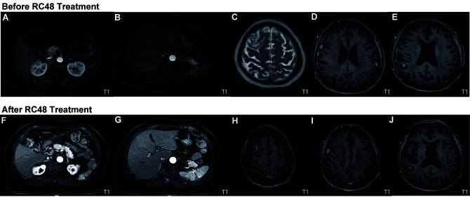
Figure 3.
Comparison of MRI prior to (June, 2021) and after (December, 2021) third-line treatment based on the newest evidence. On contrast-enhanced abdominal T1-weighted MRI, abnormal small nodular signals were observed in (A and F) segment II of the left lobe and in segment VI of the right lobe of the liver, and (B and G) a wedge-shaped abnormal signal area was observed in the subcapsular of the spleen (10×13 mm2). Contrast-enhanced brain MRI indicated (Cand H) multiple brain metastases in the bilateral frontal lobes, (D and I) leptomeninges and (E and J) right parietal lobe. The results indicated that there was no obvious progression after administration of RC48 and the imaging findings were rated as stable disease.

