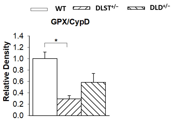
Figure 9.
Western blot analysis and relative density changes for glutathione peroxidase (GPX), normalized for cyclophilin D (CypD) protein expression, in brain mitochondria isolated from the wild-type and KGDHc-subunit-deficient mice. White bars: wild-type (WT); bars with left diagonal stripes: dihydrolipoyl succinyltransferase mutation (DLST+/−); bars with right diagonal stripes: dihydrolipoyl dehydrogenase mutation (DLD+/−). Results are expressed as means of the relative densities ± S.E.M. (N = 3–4). Statistically significant differences are indicated by asterisks; * p < 0.05.

