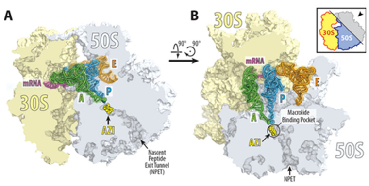
Figure 2.
Structure of azithromycin in complex with the 70S ribosome carrying A-, P-, and E-site tRNAs. (A,B) Location of the ribosome-bound azithromycin (yellow) in the macrolide binding pocket at the entrance to the nascent peptide exit tunnel (NPET) of the 70S ribosome relative to tRNAs viewed as cross-cut sections through the ribosome. The 30S subunit is shown in light yellow, the 50S subunit is in light blue, the mRNA is in magenta, and the A-, P-, and E-site tRNAs are colored green, dark blue, and orange, respectively. The phenylalanyl and formyl-methionyl moieties of the A- and P-site tRNAs are shown as spheres [15].

