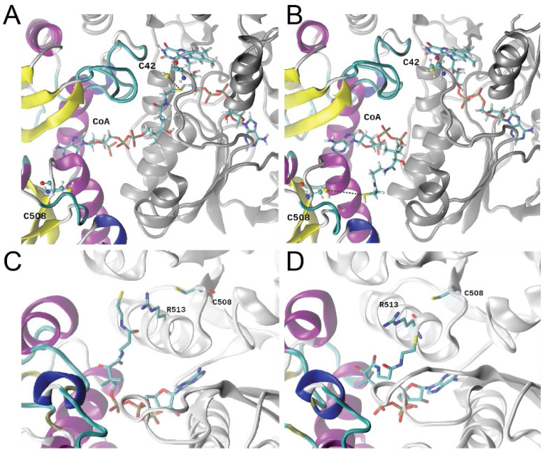
Figure 6.
Representative snapshots of stable conformations of CoA relative to C508, obtained from 1.4 µs MD simulations. (A) Starting “extended” CoA conformation, protomer B; (B) representative “bent” CoA conformation, protomer B. The dimer backbone is depicted in the ribbon, with the CDR domain of one protomer colored based on a secondary structure and the rhodanese domain from the adjacent subunit shaded silver. All residues are depicted with sticks. The position of the pantothenate arm is slightly different in replica 1 (C) vs. replica 2 (D). approaching C508 from opposite sides of the R513 side chain (shown in sphere and stick).

