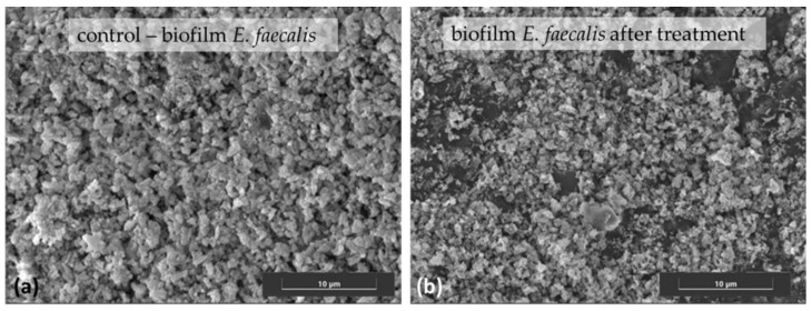
Figure 2.
SEM images of E. faecalis on urinary catheter surface (a) before and (b) after nitroxoline treatment, showing the areas of biofilm destruction. Samples were coated with Pd before analysis on a Hitachi S–3600N Scanning Electron Microscope. Image taken at 13.3 k magnification (scale bar represents 10 μm).

