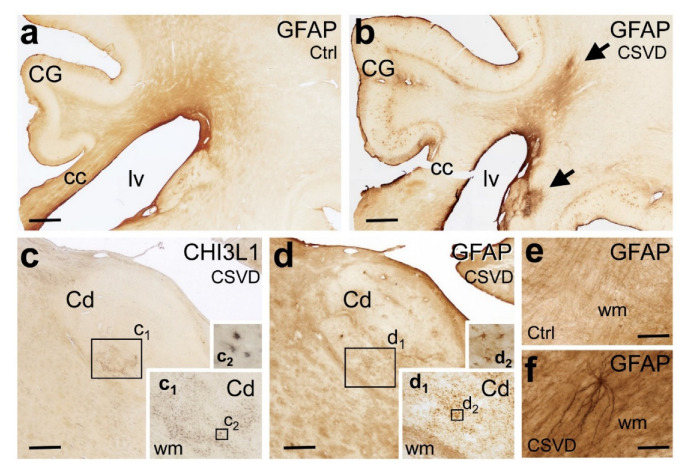
Figure 6.
Immunohistochemical demonstration of the astroglial markers glial acid fibrillary protein (GFAP) and chitinase 3-like 1 (CHI3L1) in 100-µm-thick mid-hemisphere sections in CSVD and the controls. (a,b) CSVD case (b; Case # 1) with MRI-visible white matter hyperintensities (WMH) displays stronger GFAP immunoreactivity in the white matter (arrows) than the case with acute stroke but only minor subcortical CSVD and no WMH (a; Case # 12). (c,d) CSVD case (c,d; Case # 7) with clusters of CHI3L1-positive astrocytes in the caudate nucleus (Cd) in the absence of immunoreactive astrocytes in the neighbouring white matter. The density of GFAP-expressing reactive astrocytes is also high in the Cd, whereas the surrounding white matter tracts contain bundles of GFAP-positive fibrillary astrocytic processes. Boxed areas in (c) with CHI3L1-positive glial cell clusters and in (d) with GFAP-positive glial cell clusters are shown at higher magnification in the insets (c1,c2) and in the insets (d1,d2), respectively. (e,f) At higher magnification, the periventricular white matter of CSVD cases (f; Case # 5) presents with a much higher density of GFAP-positive fibrillary astrocytic processes than controls (e; Case # 9). Cc: corpus callosum, Cd: caudate nucleus, CG: cingulate gyrus, CHI3L1: chitinase-3-like protein 1, CSVD: cerebral small vessel disease, Ctrl: control, GFAP: glial fibrillary acidic protein, lv: lateral ventricle and WMH: white matter hyperintensities. Scale bars: 1000 µm (a–d) and 100 µm (e,f).

