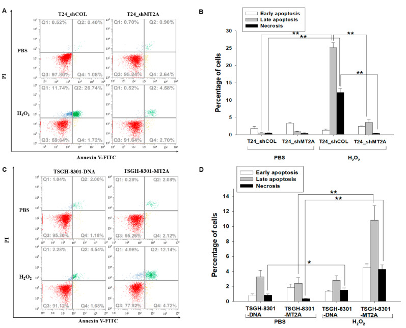
Figure 3.
MT2A enhances H2O2 treatment-induced cell apoptosis in bladder carcinoma cells. Cell apoptosis was determined by the association of annexin V-FITC with PI staining. The fluorescence intensity of mock-knockdown (T24_shCOL) and MT2A knockdown (T24_shMT2A) cells after 500 μM of H2O2 treatment for 3 h was determined by flow cytometry (A). The quantitative data were presented as the percentage of early apoptosis, late apoptosis and necrosis of cells after treatments as indicated in T24 cells (B). Flow cytometry was used to determine the fluorescence intensity of mock-overexpressed TSGH-8301 (TSGH-8301_DNA) and MT2A-overexpresssed TSGH-8301 (TSGH-8301-MT2A) after 500 μM of H2O2 treatment for 3 h (C). The quantitative data were presented as the percentage of the early apoptosis, late apoptosis and necrosis of cells after treatments as indicated in TSGH-8301 cells (D). Fluorescence intensity of the annexin V-FITC is plotted on the x-axis, and the PI is plotted on the y-axis. * p < 0.05; ** p < 0.01.

