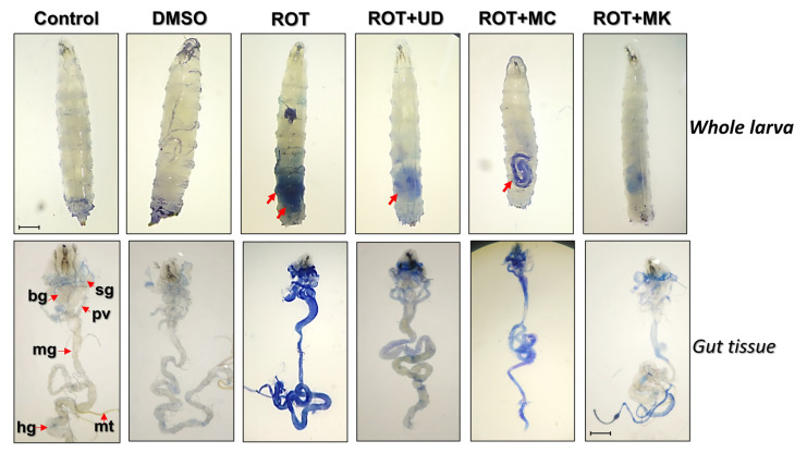
Figure 6.
Dye exclusion test through trypan blue staining in third instar larvae exposed to rotenone and cotreated with UD, MC, and MK, as shown in the upper panels. The lower panels show dissected third instar larvae stained with trypan blue. Seventy-two hour (±2 h) old larvae (early third instar) of D. melanogaster (Oregon R+) were exposed to ROT 500 µM alone or in combination with UD, MC, and MK for 48 h. Arrows of the upper panel show cytotoxicities in the whole larvae through trypan blue staining. Note: bg= brain ganglia, sg= salivary glands, pv= proventriculus, mg= midgut, mt= malpighian tubules, and hg = hind gut. The bar represents 100 μm. ROT= rotenone; UD = Urtica dioica, MC = Matricaria chamomilla and MK = Murraya koenigii.

