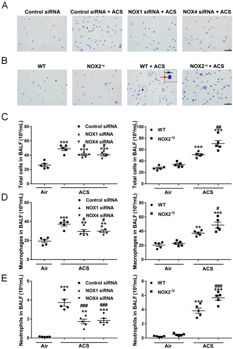
Figure 4.
Knockdown of NOX1 or NOX4 in mice attenuated ACS induced inflammatory cell accumulation in BALF. Mice were exposed to CS or filtered air for 1 h, and BALF was collected at 6 h. Cells isolated from BALF were counted with a hemocytometer and stained with Giemsa. (A) Representative images of cells in BALF from ACS exposed mice transfected with control siRNA, NOX1 siRNA, or NOX4 siRNA. (B) Representative images of cells in BALF from ACS exposed WT mice and NOX2−/y mice. (C) Total cell counts in BALF in ACS exposed mice silenced or knocked out of NOX1, NOX2, or NOX4 expression. (D) The number of macrophages (red arrowhead) in ACS exposed mice silenced or knocked out of NOX1, NOX2, or NOX4 expression. (E) The number of neutrophils (black arrowhead) in BALF in ACS exposed mice silenced or knocked out of NOX1, NOX2, or NOX4 expression. All data are presented as Mean ± SEM, n = 5. Scale bar, 50 µm. ** p < 0.01, *** p < 0.001 vs. air exposed control group. # p < 0.05, ## p < 0.01, ### p < 0.001 vs. ACS exposed group.

