Figure 5.
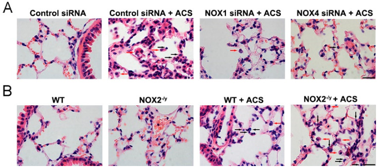
Knockdown of NOX1 or NOX4 in mice attenuated ACS induced lung inflammation. Mice were exposed to CS or filtered air for 1 h and harvested at 6 h. Lung tissue sections were stained with H&E, and the data indicate an increase in the numbers of macrophages (red arrowheads) and neutrophils (black arrowheads) infiltrating alveolar spaces in response to ACS in control siRNA-transfected or WT control mice. (A) Representative H&E images indicating that ACS exposed mice silenced of NOX1 or NOX4 expression had reduced inflammatory cell infiltration compared to control siRNA-transfected mice. (B) Representative H&E images indicating that ACS exposed NOX2−/y mice had increased inflammatory cell infiltration compared to WT control mice. Scale bar, 20 µm.

