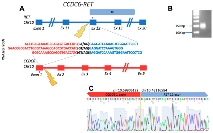
Figure 1.
Schematic representation of the CCDC6-RET fusion transcript identified: (A) gene structures upstream and downstream of fusion site; (B) electropherogram of RT-PCR product obtained with primers complementary to the fusion moieties. The deduced PCR product size is 143 bp long; (C) Sanger sequencing of RT-PCR product confirms the fusion of exon 1 of CCDC6 with exon 12 of RET. Black arrows denote position of PCR primers. TK, tyrosine kinase domain within the structure of RET.

