Figure 3.
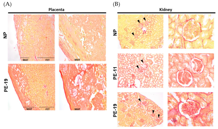
Collagen deposition in placenta and kidney. Representative pictures of (A) placenta (left) and (B) kidney (right) slices stained with Sirius Red dye. Images are organized into two columns per animal group with magnifications as follows: 4× (left column) and 10× (right column) for placenta, and 10× (left column) and 40× (right column) for kidney samples. In the kidney, glomeruli are indicated by black arrowheads. NP: normal pregnant rats; PE-11: preeclamptic rats at GD11; PE-19: preeclamptic rats at GD19; MAT: maternal portion of the placenta; FET: fetal portion of the placenta.

