Figure 8.
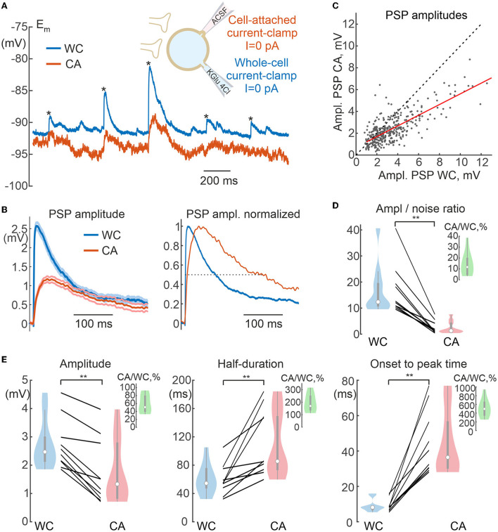
Spontaneous postsynaptic potentials in cell-attached and whole-cell recordings. (A) Scheme of recordings. Spontaneous postsynaptic potentials (sPSPs) are recorded using CA and WC electrodes from same neuron at resting membrane potential. WC electrode contains a low-chloride solution (4 mM), therefore most sPSPs are glutamatergic in nature. (B) Averaged sPSPs in WC and CA recordings with a common voltage scale (left panel) and normalized to peak amplitude (right panel), dashed line is placed at half-amplitude. The resting membrane potential values are subtracted. (C) Relationship between amplitudes of sPSPs in the WC and CA configurations for the cell shown in (A). Each dot represents an individual sPSP. The red line shows a linear fit. (D) Amplitude to noise ratio for sPSPs in WC and CA recordings. The inset shows CA/WC transfer coefficient values. (E) Parameters of sPSPs in paired WC and CA recordings: amplitude of sPSPs (left), half-duration (middle), and time from onset to peak (right). The insets in green show the values of the respective CA/WC transfer coefficients. (C,D) Pooled data from 10 pyramidal cells in the L5 somatosensory cortex of the mouse. **P < 0.01.

