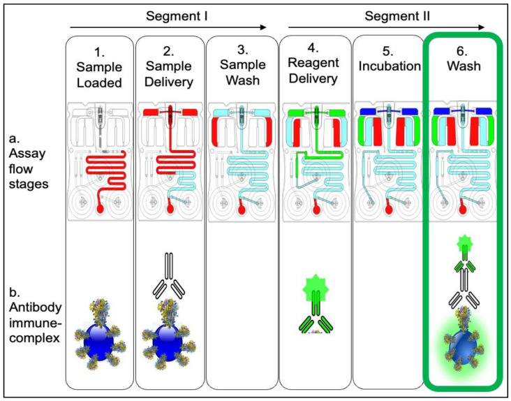
Figure 2.
The COVID-19 antibody assay sequence. Step 1 shows the sample (+/− antibody) loaded to the cartridge input port, followed by sample delivery over the bead array through buffer flow via right blister (Step 2) and finished with a wash step (Step 3). Step 4 shows introduction of the antibody detection reagent conjugated to Alexa Fluor 488 that is eluted from the reagent pad over the bead array, followed by final incubation (Step 5) and final wash (Step 6) steps. In the presence of SARS-CoV-2 IgG1 antibody in the sample, the postassay completion image shows the antibody capture beads fluorescing as a result of the antibody immune complex formation (Step 6b).

