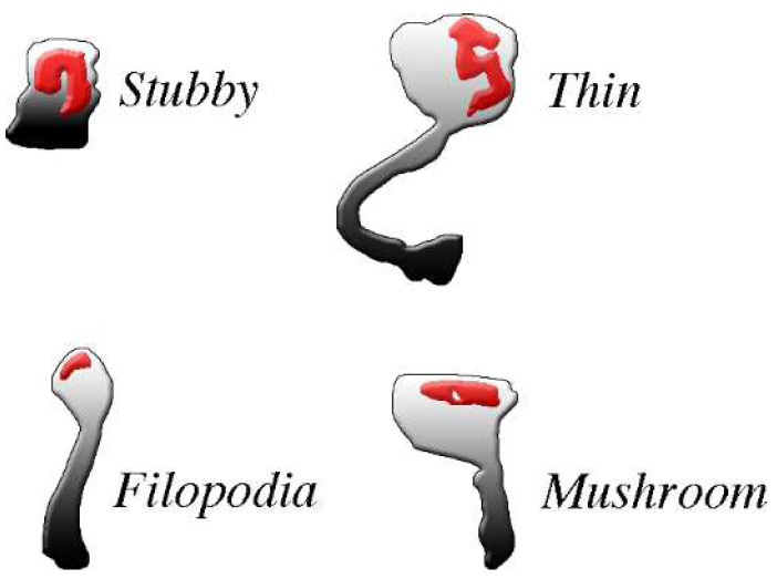
Figure 2.
The four common types of post-synaptic spines are clearly different from each other. The density and shape of post-synaptic spines, abundant in excitatory neurons, change profoundly during physiological and pharmacological events. For example, changes occur during spine generation, by continuous turnover with regeneration, and by conversion of one type of spine into another. All spines exhibit abundance of PSD (red) distributed within the body in the proximity of the plasma membrane where pre-synaptic messages are received. PSDs are widely composed by adhesion molecules bound by scattered receptors, enzymes and at least some scaffolding proteins. Most PSD-bound proteins are critical for post-synaptic responses. Spines exhibit distinct shapes: stubbles do not have long necks, thus their responses are similar to those of the flat post-synapses; filopodia, often active in groups, are long but thin, with very small heads; mushroom and thin spines exhibit relative large heads, with flat and round top surfaces, respectively, connected to their dendritic fibers by long or very long necks. Their activities tend therefore to operate independently, with limited interactions with their dendritic fiber. Permission for this Figure, a fraction of Figure 2 of [36], has been obtained from Frontiers in Neuroscience.

