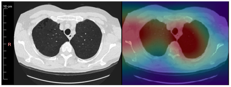
Figure 5.
An inspiration chest CT slice of a 65-year-old male patient with mild emphysema is visualized in the lung window level 1500/−500 and reconstructed using medium smooth kernel. Left: CT scan with minimum intensity projection of slab thickness 5 mm Right: A saliency map generated by a convolution of an autoencoder that is overlayed onto the CT image.

