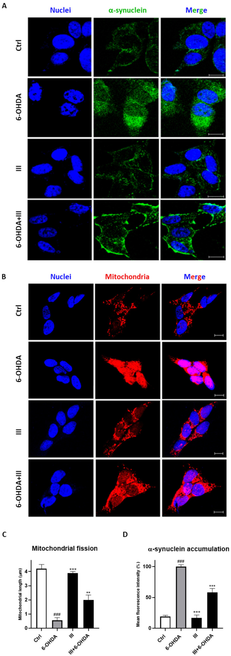
Figure 7.
Effect of fraction III (6 µg/mL) on α-synuclein accumulation (A,D) and mitochondrial fission (B,C), evaluated by anti-α-synuclein antibody and Mitotracker Red CMXRos, respectively, via confocal microscopy on SH-SY5Y cells treated with 6-OHDA (50 µM). Quantitative analysis was conducted on ImageJ, version 1.47. Data are expressed as mitochondrial length (µm) (C) or mean fluorescence intensity (D). Scale bars: 10 μm. N ≥ 30. Results are showed as mean ± standard deviation (SD) from three independent experiments. ### denotes p < 0.001 vs. Ctrl; **, *** denote p < 0.01 and p < 0.001 vs. 6-OHDA.

